Showing posts with label armature fabrication. Show all posts

Monday, August 22, 2016

Ecorche Armature Design

Available here is my design for the Ecorche Armature
I've assembled detailed plans and explanation for building your own Ecorche Human Anatomy Armature.
I currently teach Artistic Anatomy- Drawing the Human figure at the Woodstock School of Art in Woodstock, New York for the last six years. I have also taught a class in Sculptural Ecorche.
My first Ecorche was with Eliot Goldfinger at the New York Academy of Art in New York.
I've had quite a bit of experience in building these Ecorche armatures as I've fabricated about ten of these and have perfected my design on each succesive one that I build.
For those interested in purchasing one I have a few available through this website as  well as detailed plans for how to fabricate your own Ecorche armature.
They can be obtained here:

Full sized Armature with base:  $200.00 plus shipping.


Complete plans and instructions on how to build an anatomical armature
download pdf. file
$ 40.00


Tuesday, January 12, 2016

Ecorche and Artistic Anatomy at the Woodstock School of Art


Ecorche and Artistic Anatomy at the Woodstock School of Art


Ecorche Armature


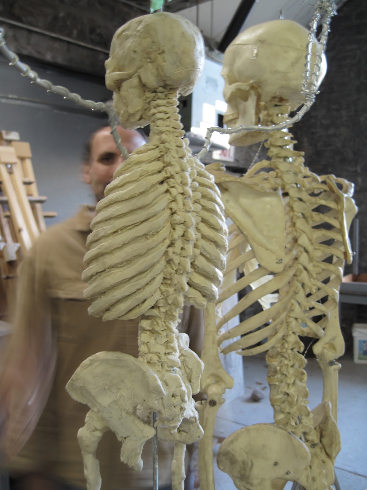
Here's the basic armature I created for my Ecorche class at the Woodstock School of Art .
Based on a single pole concept rather than a side mount, I felt that the side mounted galvanized pipe occluded too much area so I opted to hang the armature off a single thin threaded steel rod which was made rigid through wing nut compression. The base had a countersunk void to accommodate a locking nut. I spent alot of time poking through the plumbing fixture bins at the hardware store. This is actually the second prototype of my ecorche, which  was later improved upon. Hands and feet were added later, fab'd from smaller gauge wire and the pelvis and rib cage was refined to avoid any metal poking through the clay skin. This was especially evident around the lumbar and sacral area as I had to attach the leg wire assembly to this  junction and have it still appear delicate. More pixs to come.


Sunday, March 31, 2013

Half Skeleton Muscle Demo Prop


I've been using this armature I made from an articulated plastic skeleton to demonstrate muscle origin and insertion. It's a old somso cast I believe, so the detailing is better than a Bucky. After extensive use, it started to fall apart so I tweaked the armature using 3/8" galvanized pipe and a 1/2" conversion coupling to 1/2" nipple and phlange.so now it's ready to go.
This time I'm going to use the kleen klay unsulpherated terra cotta color.... it's more life like.


Monday, February 18, 2013

Frontalis muscle


The frontalis muscle, also known as the occipitofrontalis or epicranius, is thin, of a quadrilateral form, and intimately adherent to the superficial fascia. It is broader than the Occipitalis and its fibers are longer and paler in color. It is located on the front of the head.
It has no bony attachments.
Its medial fibers are continuous with those of the Procerus; its immediate fibers blend with the Corrugator and Orbicularis oculi, thus attached to the skin of the eyebrows; and its lateral fibers are also blended with the latter muscle over the zygomatic process of the frontal bone.
In the eyebrows, its primary function is to lift them (thus opposing the orbital portion of the orbicularis), especially when looking up. It also acts when a view is too distant or dim.
From these attachments the fibers are directed upward, and join the galea aponeurotica below the coronal suture.
The medial margins of the Frontales are joined together for some distance above the root of the nose; but between the Occipitales there is a considerable, though variable, interval, occupied by the galea aponeurotica.
It could be part of occipitofrontalis muscle.

File:Frontalis muscle animation small.gif

Levator Labii Superioris and Corrugator muscles


Superior to the mouth are the Levator muscles.
Levator Anguli Oris( lying underneath the Levator Labii Superioris)
The Levator Labii Superioris has a medial portion which continues superior toward the corner of the eye.
The medial fiber is known as the Levator Labii Superioris Alaeque nasi muscle.


The levator labii superioris (or quadratus labii superioris) is a muscle of the human body used in facial expression. It is a broad sheet, the origin of which extends from the side of the nose to the zygomatic bone.
Its medial fibers form the angular head, which arises by a pointed extremity from the upper part of the frontal process of the maxilla and passing obliquely downward and lateralward divides into two slips.
One of these is inserted into the greater alar cartilage and skin of the nose; the other is prolonged into the lateral part of the upper lip, blending with the infraorbital head and with the Orbicularis oris.
The intermediate portion or infraorbital head arises from the lower margin of the orbit immediately above the infraorbital foramen, some of its fibers being attached to the maxilla, others to the zygomatic bone.
Its fibers converge, to be inserted into the muscular substance of the upper lip between the angular head and the Caninus.
The lateral fibers, forming the zygomatic head, arise from the malar surface of the zygomatic bone immediately behind the zygomaticomaxillary suture and pass downward and medialward to the upper lip.
The two Zygomaticus,minor and major,extend from the lateral and posterior portion of the Zygomatic bone( far back on the cheek and attach on the oris and lip tissue.
There are two sections of the Obicularis Oris muscle, i.s., the palpebral section (eyelids) and orbital section(sheathing out the socket anteriorly.
Medial and extending from the bridge of the nose lateraly toward either side is the Nasal muscle


 Just above the obicularis muscle is a angled fibrous section of muscle that covers the glabellar swelling.
It is known as the Corrugator muscle.
The Corrugator supercilii is a small, narrow, pyramidal muscle, placed at the medial end of the eyebrow, beneath the Frontalis and just above Orbicularis oculi.
It arises from the medial end of the superciliary arch; and its fibers pass upward and laterally, between the palpebral and orbital portions of the Orbicularis oculi, and are inserted into the deep surface of the skin, above the middle of the orbital arch.
The Corrugator draws the eyebrow downward and medially, producing the vertical wrinkles of the forehead. It is the “frowning” muscle, and may be regarded as the principal muscle in the expression of suffering]. It also contracts in order to prevent high sun glare, pulling the eyebrows toward the bridge of the nose, making a roof over the area above the middle corner of the eye and typical forehead furrows.
File:Corrugator supercilii muscle animation small.gif

Sunday, February 10, 2013

Muscles of the Head


This is a compendium to the Ecorche survey.
In this installment I'll be presenting an overview of the muscles of the skull and their
influences on surface anatomy.function, and expression.


Edward Lanteri, in the classic  Modeling and Sculpting the Human Figure, divides the muscles of the head into seven groups.Masticatory, Epicranial, Auricular, Ocular, Palpebral, Nasal, and Labial.. Palpebral refers to the palpebral fissure or the "eye opening" which together with the Ocular,Nasal and Labial and with some limited movement being contributed by the frontalis of the epicranial group, compromise the muscles of expression.The Masticatory, or muscles of chewing, include, the Temporal, the Masseter, the Internal Pterygoid, the External Pterygoid, and the Buccinator muscles.For the student of Drawing and Sculpture, the moving muscles of the head can thus be further simplified to the muscles of chewing/nourishment acquisition which are primarily side plane, and the muscles of expression which are primarily front plane.


For further reading a detailed illustrated course text is avaiable here:

Constructing the Skeleton


The first step in modeling the skeleton onto the armature is of course to choose thr right kind of clay substrate for the sculpture.  Ecorches are modeled in non drying plastilina to facilitate the evolution of the modeling process and in addition to prevent details from drying and chipping away from the armature as would be a problem using wet clay, although I'm sure Houdon's majestic Ecorche he fabricated was in natural terracotta.After much experimentation and numerous Ecorches, we have chosen the non sulfurated Kleen Klay and for aesthetic reasons have chose to model the skeleton in ivory colored clay and apply the muscles in the terra cotta color.


.

The Armature


The Ecorche armature

Central to the building of the Ecorche as it is to any sculpture is the internal foundation onto which the clay conforsa and acquires its stability. The armature for the Ecorche is designed to be embedded inside the skeleton and to support the delicate framework onto which the muscles are built.After much experimentation, a sturdy and lightweight armature was constructed from galvanized 3/4"pipe nipples, couplings and phlange along with various gauges of aluminum armature wire , threaded steel rod, fittings and hardware.
A sturdy plinth was constructed from 7/8" Luan ,pine planks underneath to minimize warping and a pine pedestal through which lock nuts and armature wire could be inserted.


A detailed instructional ebooklet on how to build an ecorche armature is avaiable here: